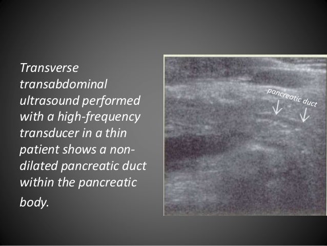
The diameter of the main pancreatic duct is a commonly assessed parameter in imaging. 41394size03 courtesy ashley davidoff md code pancreas size normal anatomy imaging radiology usscan size of the pancreatic duct the average maximum diameter of the main pancreatic duct autopsy series was 29 mm 50yrs and 35 mm 50years.

High Serum Igg4 Concentrations In Patients With Sclerosing
Normal pancreatic body measurement. Push abdomen out to make a beer belly 1. Its normal reported value ranges between 1 35 mm 58. Ultrasound images were obtained by sagittal scanning at the epigastrium and the long axis and the short axis of the oval cross section were measured. The diameter of duct can increase with inspiration 3. Adjuncts to improve visualization. The duct diameter is greatest at the head and neck region and is slightly narrower towards the body and tail.
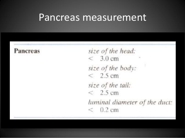
The duct in the regions of the headneck and body was measured in the transverseoblique planes. The realtime ultrasound images of the head of the pancreas in 93 children and those of the body of the pancreas in 143 children were analyzed to determine the normal size. In a prospective ultrasonic study of the pancreatic duct 233 sonograms were obtained from 49 normal subjects. In addition to the above a right subcostal approach with the transducer angled medially may be useful 1. The problems of optimal demonstration of the pancreas in ct and the possible causes of misinterpretation of the pancreatic axial tomography are considered. The size of the normal pancreas was found to be up to 30 cm for the head 25 cm for the neck and body and 20 cm for the tail.
Anterior subxiphoid approach with the left lobe of the liver as an acoustic window.


